Dry as a Bone: What to Expect if Your Dog Has Dry Eye Disease
Disclaimer: This article is meant to be informative and was not written by a veterinarian. If you are at all concerned about your dog’s eyes, or its health in general, you should discuss your concerns with your veterinarian.
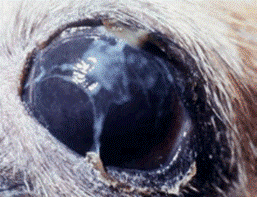
Have you ever gotten itchy eyes from staring at your laptop for too long (maybe from reading a really interesting blog post)? This is likely because you were not blinking enough and your eyes became too dry. People, and dogs, with dry eye disease feel like this all the time. This disease is actually quite similar across species, but one main difference is that people can tell their doctor when their eyes are slightly itchy. Dogs can’t. This means that disease is caught much earlier in people than it is in dogs. Dogs usually don’t get taken to the veterinarian until they have more severe signs of dry eye disease, such as yellow discharge (Figure 1), or sticky yellow goo, stuck to their eyes or eyelids. Or, they will develop cloudy, blue tinted eyes. Both of these are classic signs of inflammation of the front of the eye. This inflammation can be painful, so the dog may also squint, or not open its eyes all the way.
Figure 1. Yellow Discharge in a Dog Eye. Yellow discharge (the yellow strands stretching across this dog’s eye) is a classic sign of inflammation of the front of the eye. Dogs with this discharge should be looked at by a veterinarian as soon as possible. This image and more information on dry eye disease in dogs can be found here.
I am sure you are now wondering “what are the subtle signs of dry eye disease” so that you can watch out for them in your own pets. One of the most common early signs is that they will blink more than normal. Dogs with dry eye disease have a small volume of tears. So, they will blink more to move that small amount of tears over their eye to increase their comfort. Another subtle sign is light sensitivity which is when dogs will avoid high light areas or they will be uncomfortable opening their eyes all the way in high light environments. Squinting can also be an early sign of dry eye disease because their eyes are uncomfortable.
Figure 2. Schirmer Tear Test in a Dog. The test strip is placed under the lower eyelid. As the tears are absorbed by the paper, it turns blue. More information on dry eye disease in dogs and how to diagnose it can be found here.
If you are concerned about your dog’s eyes, you should bring it to your veterinarian as soon as you can. Once your dog is at the veterinarian, the doctor will perform some tests to determine what is wrong with its eyes. These tests will include a physical exam and an ocular exam. The ocular exam will include looking at your dog’s eye with a slit lamp, which is a handheld microscope that can be used by the veterinarian to see smaller details in the eye. The veterinarian will also perform a Schirmer tear test (Figure 2). In this test, a small piece of paper is placed underneath the dog’s eyelid to determine how much tears are on its eye. This test is very helpful because patients with dry eye disease will have small tear volumes collected with this test. Next, the veterinarian will place a yellow dye called fluorescein onto the surface of the eye. This dye will allow the veterinarian to see if there are any abnormalities to the surface of the eye. In this test, the dye will stick to areas that are abnormal, leaving a region of bright yellow stain on the eye (Figure 3). Once these tests are done, the veterinarian will use the results to determine if your dog has dry eye disease or not.
Figure 3. Fluorescein Staining of a Dog's Eye. The bright yellow region on this dog’s eye indicates that the surface of the eye is not normal in that area. More information on this stain and on how it sticks to abnormal regions of the eye can be found here.
Your veterinarian has diagnosed your dog with dry eye disease, or keratoconjunctivitis sicca (KCS). But what does that mean? Essentially, this means that your dog does not have enough tears on its eyes which caused inflammation of the cornea (the clear window at the front of the eye) and its surrounding tissues (Figure 4). This inflammation is what causes the severe signs mentioned above. For example, when the cornea is inflamed, it will turn bluish and cloudy. This is because it will fill with fluid. This increased fluid retention makes the cornea thicker, which decreases the clarity of the tissue. The conjunctiva, the tissue surrounding the cornea, is also affected in dogs with dry eye disease (Figure 4). This tissue will swell, or thicken, and become redder. However, since this tissue is mostly underneath the eyelids in dogs, it can be hard to see.
Figure 4. Eye Anatomy of the Dog. This diagram shows eye of a dog, including the cornea and the conjunctiva. The conjunctiva is the tissue that surrounds the cornea. This diagram also shows what happens when this tissue is inflamed, like it is in dogs with dry eye disease. As shown here, inflamed conjunctiva is thicker, or swollen, and redder. This diagram and more information on conjunctivitis in dogs can be found here.
But why don’t these dogs have enough tears? They don’t have enough tears because they cannot make them. The watery part of tears (Figure 5) is made by the lacrimal glands. In dogs with KCS, these glands are attacked by the animal’s own immune system. When the animal’s immune system attacks these glands, they are destroyed, which means that they cannot do their normal job. This disease can develop slowly because this destruction takes time. As the immune system attacks the gland, small pieces of the gland are destroyed and the amount of tears they produce slowly decreases.
Figure 5. Makeup of Tears. Tears have three components. The mucin layer which contains the sugar molecules that hold the watery part of the tears onto the surface of the eye. The aqueous layer, or the watery part of the tears, makes up the majority of the tears. The outermost layer is the lipid layer. This layer prevents the evaporation or loss of the watery layer. Most dogs with dry eye disease do not have enough of the watery part of the tears. This image shows a human eye, but the tears of dogs are the same.
Because this destruction is caused by the immune system, it can be treated with drugs that stop the immune system. For dogs with KCS, these drugs are usually given as eye drops to decrease the attack on the glands and to treat the inflammation of the cornea and conjunctiva. These medications help decrease the destruction of the glands. However, they cannot heal the destruction that the immune system already caused. What this means is that these dogs will likely never return to their normal level of tear production. Because of this, these dogs are often given tear replacing eye drops to help increase their tear volume. Together, these medications can dramatically increase the comfort of dogs with KCS.
Overall, dry eye disease is manageable in dogs. But, it can become serious if it is not found early. Checking your animals for subtle signs of disease and discussing any concerns about your dog’s health with your veterinarian can help identify the disease earlier and enable you to maximize your animal’s comfort.
Erin Hisey is a second year PhD student in the Integrative Pathobiology Graduate Group. She is studying a new treatment for dry eye disease in both humans and animals. She is also a veterinary student at the University of California, Davis, School of Veterinary Medicine.
For more content from the UC Davis science communication group, “Science Says,” follow us on Twitter @SciSays.


